Introduction
The surgical removal of the mandibular third molar (M3M) is a common procedure performed by oral and maxillofacial surgeons, oral surgeons, and general dental practitioners. In England, it has been estimated that more than 150,000 patients have third molar surgery each year.1 Most M3Ms that require surgical removal are usually impacted due to failure of eruption into occlusion beyond their chronological age of eruption.2 The impaction of third molars, especially in the mandible, is reported to occur in 58% of individuals, and can cause a myriad of clinical issues such as pain, swelling and infections.3 The benefits of M3M removal may include rendering the area free of symptoms or disease. However, apart from risks such as pain, swelling, bruising, bleeding, trismus, alveolar osteitis, postoperative infection, and damage to adjacent structures, which are common to all surgical extractions, M3M removal includes the risk of permanent or temporary damage to the inferior alveolar nerve (IAN) or the lingual nerve.4 IAN damage can significantly impact on quality of life (QoL) due to an altered sensation of the lip and chin area. Factors such as horizontal impactions, close radiographic proximity to the inferior alveolar canal, or patients over the age of 24 years, are associated with significantly higher risk of IAN damage.5 As such, the alternative surgical option of coronectomy, which is the sectioning of the crown from the tooth and deliberate retention of the roots, can be offered to high-risk cases to avoid IAN damage.6 This case report discusses a patient who underwent the alternative surgical option of coronectomy for her left M3M – which had a high risk of IAN damage – instead of a surgical removal.
Case presentation
A 36-year-old female was referred by her general dental practitioner to the oral surgery department at the Royal London Dental Hospital regarding her left M3M. The patient complained of recurrent throbbing and aching localised pain from the left M3M, which had occurred for over six months. The pain usually lasted for a week and had accompanying symptoms such as swelling and trismus. She managed her symptoms pharmacologically with over-the-counter analgesics (paracetamol and ibuprofen). In addition, she had been prescribed a course of antibiotics (amoxicillin, 500mg three times daily for five days) by her general dental practitioner, which helped to ease her symptoms. The patient was fit and well with no relevant medical history. She was not taking any prescribed medications and had no known drug allergies. She was a non-smoker and only consumed alcohol on rare occasions. The patient had had a mesially impacted right M3M surgically removed under local anaesthetic about six months previously, with no complications reported, and subsequently had a restoration on the lower right second molar.
Examination and investigations
On examination, there was no palpable lymphadenopathy, facial asymmetry or swelling. The patient’s temporomandibular joints and muscles of mastication had no abnormality detected and were functioning within normal values. There was no abnormality detected in her soft tissues (palate, tongue, floor of mouth, buccal mucosa). Her left M3M was partially erupted and horizontally impacted against the left mandibular second molar, and while the surrounding operculum appeared inflamed, there was no sign of swelling or pus exudation. However, the operculum posed as a food trap, making it difficult for the patient to clean. The left maxillary third molar was over-erupted and appeared to be traumatising onto the opposing third molar’s operculum. The right M3M extraction site had healed well with mucosal coverage. An orthopantomogram (OPG) (Figure 1) that had been taken on a previous occasion displayed a horizontally impacted left M3M. It had no associated apical pathology and appeared caries free. Its roots appeared bulbous and there was slight darkening of the mesial root. The OPG also displayed bifurcation of the IAN, which was present on bilateral sides of the mandible. A cone beam computer tomography (CBCT) scan (Figures 2-4) was taken to further investigate its relationship. It displayed three fully formed roots on the left M3M: a fused mesiobuccal and mesiolingual root, and a distal root. The roots had a degree of hypercementosis that contributed to the bulbosity. The canal appeared inferior to the mesial root in its mid-third, then passed along the buccal aspect of its apex and buccal to the distal apex. Furthermore, there was a loss of cortical outline and a slight narrowing of the canal associated with the mesial root and distal apex. There was a prominent branch off the canal, which was buccal to the coronal third of the roots and exited the mandible approximately distobuccal to the crown.
Diagnosis and treatment
The diagnosis of recurrent pericoronitis was based on the history, and clinical and radiographic findings. The treatment options included no treatment, removal of the left maxillary third molar, or the removal or coronectomy of the left M3M with or without the removal of the left maxillary third molar. With information on the treatment options, along with the accompanying risks and benefits, the patient decided on the option of coronectomy of her left M3M. The patient was offered the choice of having the procedure carried out under local anaesthetic or with intravenous sedation. Once informed consent was obtained, the procedure was carried out under local anaesthetic.
Standard preparation and draping were carried out. Local anaesthetic solution (2.2ml 2% lidocaine with 1:80,000 adrenaline for IAN block and 2.2ml 4% articaine with 1:100,000 adrenaline for local infiltration) was administered. A full mucoperiosteal flap was raised with mesial and distal relieving incisions, and a conservative buccal trough was drilled using a straight surgical handpiece and fissure bur. Once the crown was sectioned and removed, the remaining enamel was removed and the roots were reduced to a height 3mm below crestal bone level. Copious amounts of saline irrigation were used, and before primary closure of the wound using 4-0 vicryl rapide suture, the roots were checked for mobility. Haemostasis was achieved and the patient was given written and verbal postoperative instructions before discharge. The procedure was carried out without any complications, and no postoperative antibiotics were prescribed.
Outcome and follow-up
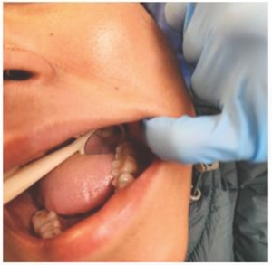
The patient was clinically reviewed at 10 and 12 weeks after the surgery at her request. She reported postoperative pain, swelling and facial asymmetry from the left mandible that lasted for two weeks. There was no report of fever, trismus, or pus exudation. The symptoms eventually subsided and she was asymptomatic at presentation. The surgical site appeared to be healing well with mucosal coverage and no signs of infection. A half OPG was taken to assess for any pathology (Figure 5). It displayed left M3M roots in situ with no evidence of enamel remaining. There was a slight radiolucency at the apices of the roots, which may be evidence of root migration from the IAN. However, the remaining root structure still appeared to be around 3mm below crestal bone level and there was no sign of pathology noted. The symptoms of pain and swelling experienced by the patient were deemed to be a typical presentation of postoperative pain and swelling instead of an infection. The patient was reviewed once more at 12 weeks, at which time she remained asymptomatic, and the surgical site was completely healed with full mucosal coverage (Figures 6 and 7). There was no report of altered sensation of the lip, chin and tongue areas.
Discussion
Indications for surgical intervention
National Institute for Health and Care Excellence (NICE) guidelines have outlined indications for the removal of third molars, which include untreatable tooth decay, abscesses, cysts or tumours, disease of the tissues around the tooth, or if the tooth impedes other surgery.7 Diseases of the tissues around the tooth include recurrent episodes of pericoronitis, which is the swelling and infection of the gingival cuff around a partially erupted tooth. Thus, based on the diagnosis of recurrent pericoronitis of the left M3M, the patient met the criteria for surgical intervention. Furthermore, the horizontal impaction of the left M3M significantly increased the risk of distal caries developing in the adjacent mandibular second molar, which was evident on the contralateral side. This is supported by recent parameters of care by the Faculty of Dental Surgery (FDS) of the Royal College of Surgeons of England (RCSE).8
Radiographic investigations
A dental OPG is well recognised as a standard imaging tool to identify specific radiographic features in relation to risk of nerve injury.9 Although there are seven main radiographic signs associated with IAN damage (darkening of root, defected roots, narrowing of roots, bifid root, interruption of the cortical outline of the canal, diversion of the canal, and narrowing of the canal), the most significant features that indicate a close proximity to the IAN include darkening of the root, diversion of the canal, and interruption of the white cortical outline of the canal.8 The patient’s OPG displayed a slight darkening of the mesial root apex, which may indicate a close proximity to the IAN. Due to the complex root morphology and the bifurcation of the IAN, the clinical decision to take a CBCT scan in addition to the OPG was agreed on to further investigate the relationship between the roots and the IAN. Despite increasing support for the use of pre-operative CBCT when indications of close proximity to the IAN are observed,10,11 there remains a lack of evidence to show a reduction of risk of IAN damage.12–15 In addition, there is an increased exposure of four to 42 times the radiation of a dental OPG and an accompanying increased risk of stochastic effects.16,17 However, CBCT was indicated and justified in this case as it might alter the course of treatment. In addition, as the roots might be mobilised during a coronectomy procedure, it may be beneficial to be fully aware of the relationship between the roots and IAN to minimise risk of damage if it requires removal.
Treatment options
The average risk of IAN damage during M3M removal is reported to range from 0.35% to 8.4%.5 The surgical removal of this patient’s left M3M had an increased risk (up to 20%) of IAN damage due to its close proximity to the nerve.18 This was discussed with the patient, along with alternative treatment options such as no surgical intervention (which might run the risk of recurrence of symptoms, progression of disease or development of pathology to the adjacent second molar), the removal of the left maxillary third molar (which could remove the source of mechanical insult and reduce symptoms or pericoronitis), or coronectomy of the left M3M.8 The patient was a suitable candidate for a coronectomy procedure as the left M3M was caries free, vital with non-inflamed pulp, and she was not medically compromised. In addition, the position of the IAN did not impede the area where the crown would be sectioned. Despite the risk common to surgical extractions, coronectomy has been reported to reduce the risk of IAN damage by 84%.6 However, the procedure itself runs the risk of IAN damage, with an incidence of 1.3%. Furthermore, the risk of failure of the coronectomy procedure has been reported to be around 7%, which is due to mobility of the roots or root migration.19 There is still a debate in the literature regarding the long-term fate of the retained root due to the need for second-stage surgery for its removal along with its complications.20,21 It has been reported that up to 91% of roots migrate within six months, and the incidence of second-stage surgery was 2.2%.6,22 Thus, with the risks and benefits of the different options discussed, the patient was provided with sufficient information to make an informed decision.
Coronectomy
Coronectomy is an alternative surgical procedure to complete removal when the wisdom tooth is considered at high risk of IAN damage. It reduces the risk of IAN damage by ensuring retention of the vital roots that are in close proximity to the canal, thus avoiding direct or indirect damage to the IAN. The coronectomy technique used on the patient was adapted from the technique as described by Frafjord and Renton.23 This involved using the buccal approach and removal of buccal bone using a fissure bur down to the cemento-enamel junction, partial sectioning of the crown from the root using a fissure bur and elevated from the buccal approach, removal of all remaining enamel using a rose head bur, and closure of buccal flap over the roots with sutures. As no lingual retraction was used for this case, a small amount of tooth structure was left intact lingually to prevent iatrogenic damage to the lingual nerve from the bur during horizontal sectioning. The crown was then separated from the roots using a Coupland’s elevator.24–26 This technique may risk mobilising the roots, especially if the roots are conical, or if the patient is young and female.18 However, this patient’s roots had a degree of hypercementosis, which contributed to its bulbosity and reduced the chances of mobilisation. The removal of all remaining enamel is essential due to its inert dental structure of ectodermic origin, which prevents the attachment of gingival connective tissue to its surface, preventing complete mucosal coverage and facilitating alveolar osteitis and infection.27 Furthermore, the roots must be reduced to 3mm below crestal bone level to allow osseous formation over the roots and the preservation of its vitality.19,27
Complications
The patient experienced postoperative pain and swelling for two weeks, which was managed with over-the-counter analgesics. As there were no signs of infection such as trismus, purulent drainage, or fever, it was presumed that the patient did not experience a postoperative infection. Despite the concern of an increased risk of postoperative infection due to the deliberate retention of roots, it has been reported that there is no difference as compared to surgical removal.6 While the half OPG taken at 10 weeks post operatively displayed no pathology, radiolucency at the root apices may indicate a slight degree of migration. The reported incidence of root migration varies from 13% to 85% of cases, of which roots tend to migrate from 0 to 6mm within 24 months.6,22 However, migration of roots should not necessarily be considered a risk as migration away from the IAN can be beneficial if second-stage surgery was required. One limitation of this case includes the short-term follow-up period of three months, after which the patient may present with further complications such as root migration, eruption, or infection.
Conclusion
Coronectomy presents as a viable alternative surgical option for the removal of M3Ms that are at a high risk of IAN damage. This is due to the significant reduction of risk of IAN damage and low incidence of failure. Although definitive conclusions cannot be made about the long-term fate of the retained roots due to the lack of high-quality, long-term studies, patients may view the risk of a second surgery to be less than the high risk of IAN damage from the surgical removal.6 As such, the risks and benefits of the different treatment options should be clearly explained to patients for them to make an informed decision.


