Loading [Contrib]/a11y/accessibility-menu.js
1.
Banjar A, Al-Masri N, Akil A, Demyati A, Abed H. Aggressive pyogenic granuloma mimicking a malignant lesion:  Case report and review. JIDA. Published online February 16, 2023. doi:10.58541/001c.70263
Download all (5)
  • Figure 1. Pre-operative intra-oral photos.
  • Figure 2.
  • Figure 3. Histopathologic investigation using H and E stain.
  • Figure 4. Part of the surgical field during the removal of the lesion (A and B), extraction of the hopeless teeth (C), wound suturing (D), and part of the lesion after the excisional biopsy (E).
  • Figure 5. One week (A), three weeks (B) and one year (C) after the operation, showing good healing with no signs of infection.

Abstract

A pyogenic granuloma is a reactive hyperplastic inflammatory lesion that arises as a result of various stimuli such as low-grade local irritation, trauma, hormones, and certain medications. In the oral cavity, it is a relatively common gingival soft tissue lesion, which originates as an expansile growth of the mucous membrane. It could exhibit aggressive behaviour mimicking that of malignant lesions with different presentations. This article aims to present the case of a patient who attended the dental clinic while complaining about a large and rapidly growing intraoral mass with aggressive behaviour, which had manifested as a fast-growing lesion causing rapid expansion of the buccal and palatal bone, bleeding, and alveolar bone destruction associated with mobile teeth. The article also aims to compare this case with current reported cases of aggressive pyogenic granuloma in the literature.

Introduction

A pyogenic granuloma (PG) is a benign inflammatory hyperplastic lesion located in the skin and the mucous membrane.1 In the oral cavity, PGs have a higher propensity to present in females, especially in their second decade, during pregnancy, and after menopause.1,2 PGs are more commonly observed in the maxilla than the mandible, and in the anterior rather than the posterior region of the oral cavity.3 It appears under a microscope as a highly vascular proliferation that resembles granulation tissue.4

Numerous studies have tried to reveal the aetiology of PGs; however, the exact aetiology remains unclear.5 The commonly reported causes are local irritation and chronic trauma, which are responsible for initiating reactive lesions characterised by connective tissue proliferation.6 Other causes may include poor oral hygiene, parafunctional habits, a history of dental extraction, and overhanging dental restorations.5,7 In addition, medications such as calcium channel blockers, and anticonvulsant and immunosuppressant drugs, are known to induce gingival hyperplasia.8 Other researchers believe that PGs appear because of an infectious process or hormonal changes.9

A PG diagnosis depends on a biopsy and histopathological assessment, which help to differentiate from other common oral diseases.10 Its differential diagnosis includes haemangioma, peripheral giant cell granuloma, hyperplastic gingival inflammation, pregnancy tumour, peripheral ossifying fibroma, drug-induced gingival hyperplasia, and malignant lesions.11 However, the aggressive behaviour of a PG can be diagnostically challenging.12

The treatment of choice for a PG is an excisional biopsy.4 Other treatment modalities include cryosurgery. The use of an Nd:YAG laser, flash lamp pulsed dye laser, injection of ethanol or corticosteroid, and sodium tetradecyl sulphate sclerotherapy has also been documented.4

This article aims to present the case of a patient who attended the dental clinic while complaining about a large, rapidly growing intraoral mass with aggressive behaviour, which had manifested as an extensive loss of alveolar bone, as well as mimicking a malignant lesion. The article will also compare this case with current reported cases of aggressive PGs in the literature.

Clinical presentation

A 50-year-old male was referred by his general dentist in a private dental clinic to the Oral and Maxillofacial Surgery Department at King Fahad Hospital (KFH) in Makkah, Saudi Arabia. The referral was for a large lump that needed further evaluation and treatment. The patient’s medical history was remarkable for controlled diabetes and hypertension. His medications included long-acting insulin and rapid-acting insulin analogues for diabetes mellitus. The patient took calcium channel blockers and long-acting angiotensin-converting enzyme inhibitors for hypertension. The patient had been a smoker of two packs per day for more than 25 years. The lesion had developed for almost a month and a half before the first visit.

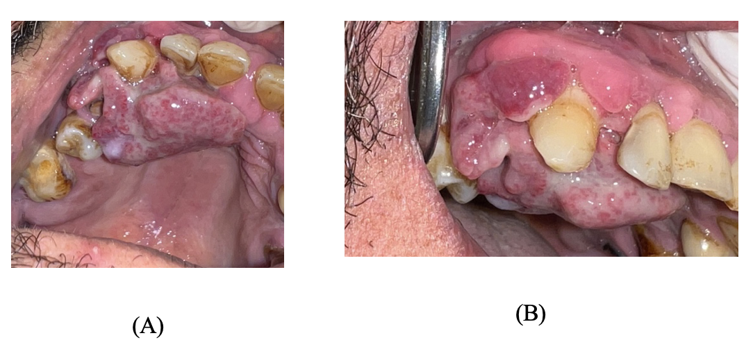
Upon extra-oral examination, no abnormalities were detected. Intra-oral examination revealed a large, exophytic, rapidly growing, soft swelling with an irregular surface extending from the upper right central incisor to the upper right first molar, causing rapid expansion of the buccal and palatal alveolar bone (Figure 1).

Figure 1
Figure 1.Pre-operative intra-oral photos.

A: Occlusal view – notice the speckled whitish-red area; B: Lateral view – notice the purplish-red buccal interdental papilla.

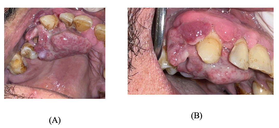
When palpated, the swelling was soft, compressible, and bled easily. Grade III mobility was associated with the upper right lateral incisor, upper right canine and upper right first premolar. In addition, grade II mobility was associated with the upper right central incisor, upper right second premolar, and upper right first molar. The patient was not in pain, but felt discomfort due to the rapid growth of the lesion. In addition, the presence of the lesion made it difficult for the patient to maintain oral hygiene, thereby favouring the lesion’s growth.

The patient denied having unexplained pain, fevers, lymphadenopathy, persistent cough, hoarseness, swallowing difficulty, loss of appetite, night sweats, or numbness in the lesion’s area, or any other warning manifestation that might raise the index of suspicion for malignancy.

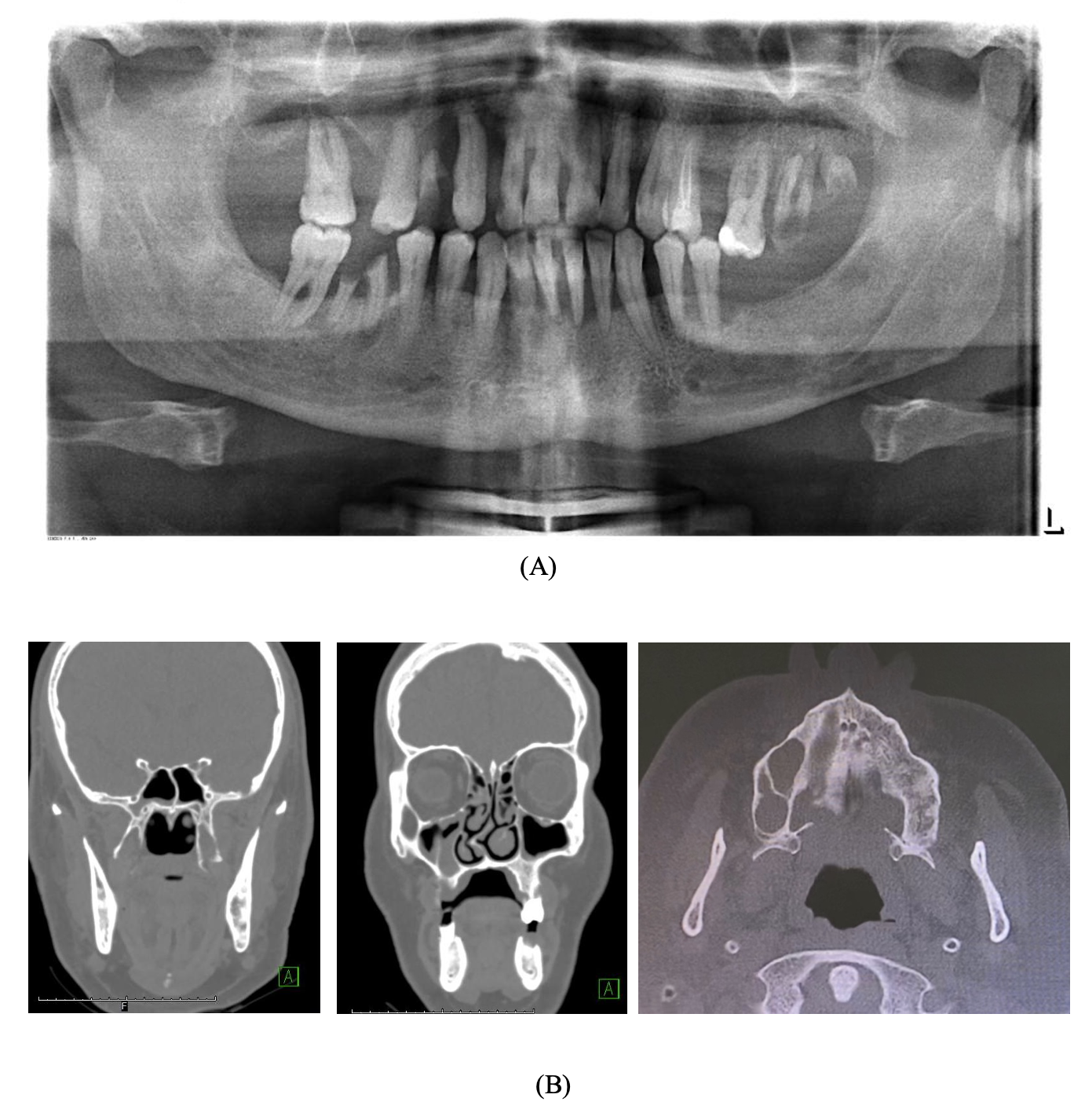
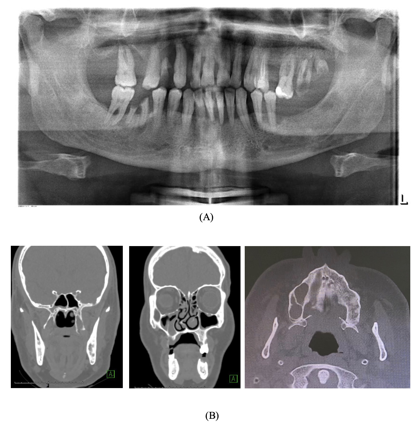
A pre-operative orthopantomogram (OPG) showed multiple non-restorable teeth in both jaws, as well as generalised, horizontal moderate to severe bone loss (Figure 2a). Pre-operative cone beam computed tomography (CBCT) showed an ill-defined lesion on the palatine process of the right maxillary and alveolar bone (Figure 2b).

Figure 2
Figure 2.

A: Pre-operative orthopantomogram showed bone destruction on the upper right side and a remaining root right first premolar and carious right lateral incisor. B: Cone beam computed tomography showed an ill-defined soft tissue mass measuring 19x20x17mm on the palatine process of the right maxillary and alveolar bone with underlying bone erosion, mucosal thickening of the maxillary, ethmoidal, frontal and sphenoid sinuses, and hypertrophied inferior conchae. The nasal septum deviated to the right.

Management

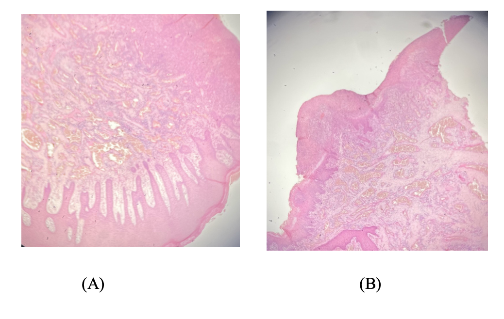
The treatment plan was discussed with the patient and began with advising him to quit smoking and participate in a smoking cessation programme. Thereafter, an incisional biopsy of the lesion was performed under local anaesthesia to detect the precise diagnosis. The surgical site was sutured using polylactic acid suture 3/0. The biopsy was sent to the histopathology laboratory, and following histopathological assessment (Figure 3), the diagnosis was of a PG.

Figure 3
Figure 3.Histopathologic investigation using H and E stain.

A: Magnification degree ×100; B: Magnification degree ×40. The histopathologic investigation revealed small capillaries arranged in a lobular fashion and vascular sinusoidal spaces. The overlying surface is ulcerated and covered by a fibrinous exudate. The endothelial cells and keratocytes of the lesion showed no atypia. No melanocytic nests were observed.

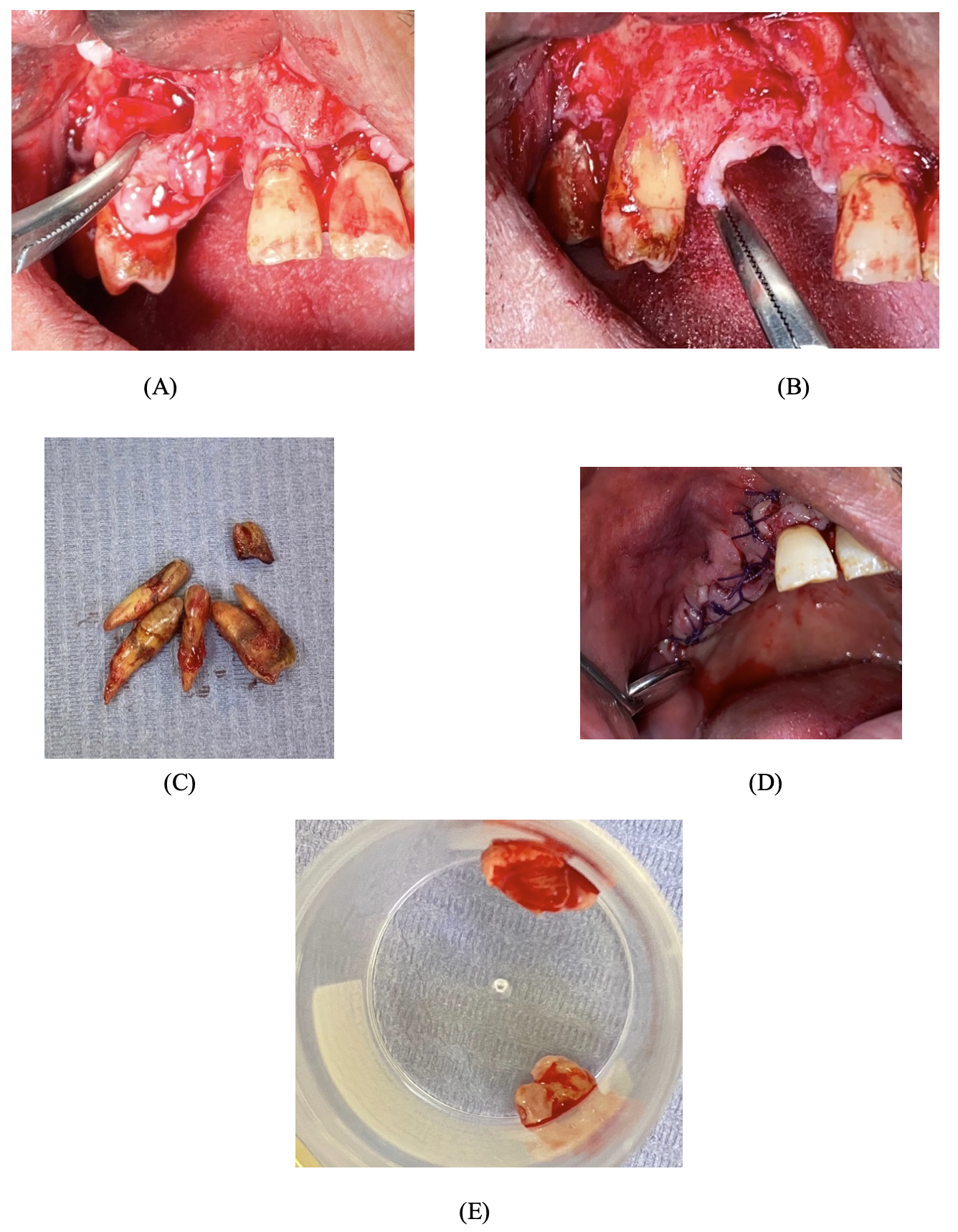
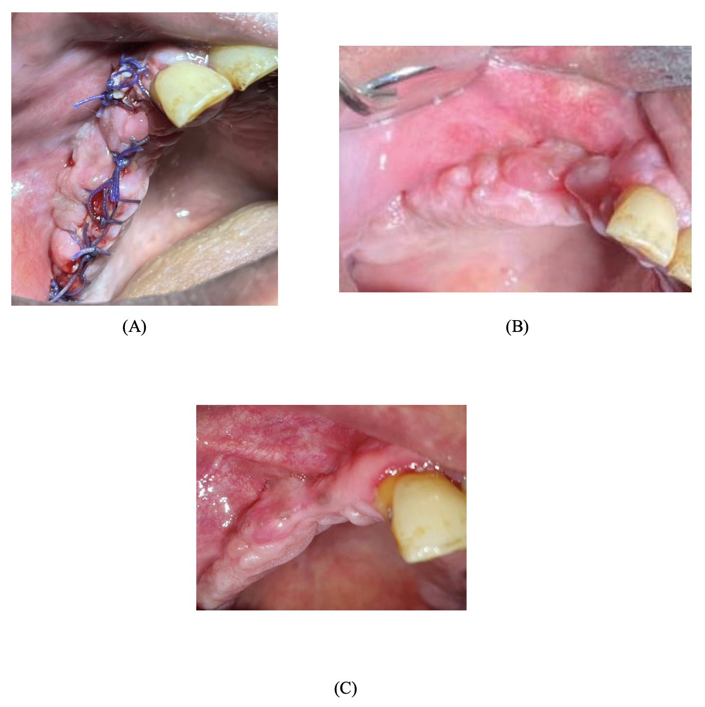
Definitive treatment included a complete excision of the lesion from the buccal and palatal aspect associated with extraction of all hopeless teeth (upper right lateral incisor, upper right canine, upper right first premolar, upper right second premolar, and upper right first molar). Curettage and alveoloplasty were performed, followed by irrigation and haemostasis achieved by suturing to promote primary healing (Figure 4).

Figure 4
Figure 4.Part of the surgical field during the removal of the lesion (A and B), extraction of the hopeless teeth (C), wound suturing (D), and part of the lesion after the excisional biopsy (E).

Postoperative instructions were given (i.e., maintain good oral hygiene and a soft diet, and avoid spitting in the first 24 hours), and antibiotic coverage of amoxicillin 500mg three times daily for seven days, as well as painkillers, was prescribed. The patient tolerated the procedure without complications and was discharged on the same day.

The patient attended his recall visits (at one week, three weeks and one year postoperatively). Clinical examination showed sufficient healing with no signs of recurrence or infection (Figure 5).

Figure 5
Figure 5.One week (A), three weeks (B) and one year (C) after the operation, showing good healing with no signs of infection.

The patient reported a satisfactory result.

Discussion and review of the literature

The current case report was conducted based on the CARE (CAse REport) Checklist developed by the Joanna Briggs Institute (JBI) at the University of Adelaide, South Australia. Ovid, Medline and Embase (1946 to 2022) were searched for relevant articles related to an aggressive PG. Clear search strategy keywords (i.e., pyogenic granuloma.​mp. or granuloma, pyogenic, aggressive granuloma.​mp., aggressive.​mp., and a combination of these keywords) were used to search the relevant articles. Three evaluators worked independently to include or exclude the relevant studies. All types of studies in the English language were included from 1946 to January 14, 2022. Only 12 articles were found to be related to aggressive PG. Ten of these were case reports (Table 1),11–20 one was a review (Table 2),3 and one was a retrospective study (Table 3).21

Table 1.Case reports included in the literature review (total = 10).
Authors Summary of the case Site of the lesion Management Conclusion
Gender Age Clinical characteristics
(Shetty et al., 2020)12 Female 13 Gingival swelling that bleeds easily while brushing, started about five months previously Anterior maxilla Excisional biopsy and extraction of affected teeth Awareness of the aggressive behaviour of pyogenic granuloma is crucial to avoid the inadvertent treatment of this reactive lesion
(Kumar and Agarwal, 2019)11 Female 11 Growth in the oral cavity located at the marginal gingiva Posterior mandible Excisional biopsy and extraction of affected teeth The appropriate management of pyogenic granuloma is related to accurate diagnosis
(Gefrerer et al., 2016)20 Female 55 Growing granuloma three weeks after dental implants were loaded with splinted porcelain-fused-to-metal crowns Posterior mandible Excisional biopsy It is important with high recurrent types of pyogenic granuloma to have a long-term follow-up
(Sharma et al., 2012)19 Female 13 Irregular gingival growth in the past two years that bleeds easily, associated with a difficulty chewing and erosion of the alveolar bone Posterior maxilla Excisional biopsy The aggressive type of pyogenic granuloma is associated with erosion of the alveolar bone, maxillary sinus invasion, mobility, and disablement of affected teeth
(Martins, Piva, da Silva, 2011)17 Female 20 Asymptomatic nodular mass of three weeks’ evolution on the buccal and palatal gingiva with bone resorption around the right lateral incisor Anterior maxilla Incisional biopsy followed by excisional biopsy Pyogenic granuloma can mimic tumours associated with extensive loss of alveolar bone
(Mubeen et al., 2011)18 Female 63 Growth in the right molar region lasting for three months Posterior mandible Excisional biopsy and antibiotic coverage of amoxicillin 500mg tid for five days Maintaining good oral hygiene and preventing recurrence of the lesions, with regular follow-up, is required when managing the aggressive type of pyogenic granuloma
(Ababneh and Al-Khateeb, 2009)16 Female 28 Massive gingival swelling with alveolar bone loss beneath the lesion Posterior mandible Excisional biopsy under general anaesthesia during pregnancy with no untoward reactions The aggressive type of pyogenic granuloma can mimic malignant tumour behavior
(Jadwat et al., 2008)15 Male 14 Overgrowth lasting for 18 months with loosening of the upper anterior maxillary teeth Anterior maxilla Excisional biopsy, antibiotic coverage and chlorhexidine
mouthwash
It is important to have an appropriate histopathological investigation to determine whether the granulomatous mass may not be a malignant lesion
(Lee and Polonowita, 2007)14 Female 37 Recurrent vascular swelling Anterior mandible Deep scaling, chlorhexidine mouthwash for two weeks and oral hygiene instruction followed by an excisional biopsy after one month It is mandatory to identify all the possible causes of the pyogenic granuloma before treatment initiation to help avoid expensive and complex treatment
(Ramirez et al., 2002)13 Female 9 Reddish, sessile swelling Posterior mandible Excisional biopsy Severe pyogenic granuloma is a common oral lesion during puberty
Table 2.Review study included in the literature review.
Authors Type of study Summary of the study Site Management Conclusion
(Jané-Salas et al., 2015)3 Review Sixteen case reports of 21 patients (i.e., 15 patients diagnosed with peripheral giant cell granuloma and five patients diagnosed with pyogenic granuloma) Mandible > maxilla Excisional biopsy and curettage, although one case involved excision with Er-YAG laser The aggressive nature of pyogenic granoluma and peripheral giant cell granuloma was associated with dental implants; however, the robustness of the evidence is unclear
Table 3.Retrospective study included in the data analyses.
Authors Type of study Aim Site Management Conclusion
(Shuster et al., 2021)21 Retrospective study To investigate the clinical and histopathological parameters of lesions mimicking peri-implantitis in 65 patients (age range 39-80 years; 62% female, 38% male) Associated with dental implants Excisional biopsy and curettage Pyogenic granuloma with aggressive behaviour was represented at 25% with a recurrence rate of 6% when associated with dental implants. The histopathological analysis is crucial to reaching a definitive diagnosis and treatment

Site of PG

Similar to the current case, five case reports reported that the site of the PG was in the maxilla.12,13,15,17,19 In the remaining five case reports, the PG was reported in the mandible.11,14,16,18,20 One review included in this article discussed PGs and found that the location of the majority of included cases was the posterior part of the mandible without bone loss.3 In addition, a retrospective study showed a site of a PG associated with a dental implant.21

Treatment modalities

In our case report the treatment of choice was an excisional biopsy under local anaesthesia as well as the extraction of hopeless teeth that were associated with the lesion. Similar to the current case, three case reports were found to treat a PG by means of an excisional biopsy.16,19,20 In one case, the treatment was oral prophylaxis followed by teeth extraction and an excisional biopsy.11 Another case used an incisional biopsy followed by an excisional biopsy after the histopathological report.17 Two other case reports used antibiotics after an excisional biopsy without teeth extraction.15,18 One case report outlined treatment of localised aggressive periodontitis combined with an excisional biopsy.15 Additionally, one of the included case reports was that of an excisional biopsy with extraction of the affected teeth without oral prophylaxis.12 What is more, chlorhexidine mouthwash was used for two weeks to decrease the lesion size, and then an excisional biopsy after one month was also reported.14 In addition, a bone trough was reported as a method of management.20 The included review in the data analyses illustrated 16 cases of PGs, with successful treatment including an excisional biopsy and curettage.3

Aggressive pyogenic granuloma

The current case presented a diabetic and hypertensive patient, who attended the dental clinic while complaining about an aggressive lesion. Our case, supported by other reported cases in the literature, showed that an aggressive PG could appear clinically as an irregular soft lesion, purple and reddish and bleeding easily, with resorption of the alveolar bone. On the other hand, the clinical picture of a PG is often characterised as an asymptomatic, painless lesion covered by a yellow fibrinous membrane that grows slowly without bone loss.10 In addition, an aggressive PG can be associated with mobile teeth in the lesion area.

The appearance of the aggressive PG in our case report is similar to that of aggressive PG developing in association with dental implants. For example, Jané-Salas et al. (2015) emphasised in their review that aggressive PG is associated with cases that include dental implants.3 Similarly, in their retrospective study, Shuster et al. (2021) found that aggressive PG was represented at 25%, with a recurrence rate of 6% when it was associated with dental implants.21

Conclusions

A PG could exhibit aggressive behaviour that mimics malignant lesion behaviour, with fast growth, rapid expansion of the buccal and palatal bone, bleeding, and alveolar bone loss associated with mobile teeth. Aggressive PGs were reported to be more commonly associated with dental implants, mimicking peri-implantitis. Histopathological assessment is essential in order to exclude any potential malignancy and help with early referral to formulate a definitive diagnosis and treatment plan.


Acknowledgement

The authors would like to acknowledge histopathology consultant Dr Nasir Al-Noor at King Faisal Hospital (Makkah, Saudi Arabia) for his contribution to this project by reporting and reading the histopathological slides.

Accepted: January 03, 2023 IST

References

1.
Mighell AJ, Robinson PA, Hume WJ. Immunolocalisation of tenascin-C in focal reactive overgrowths of oral mucosa. J Oral Pathol Med. 1996;25(4):163-169. doi:10.1111/j.1600-0714.1996.tb00214.x
Google Scholar
2.
Mohan M, Karikal A, Bhat S, Padmaja A, Thilak G. Aggressive pyogenic granuloma causing bone erosion: case report. Healtalk. 2012;4(3):13-14.
Google Scholar
3.
Jané-Salas E, Albuquerque R, Font-Muñoz A, González-Navarro B, Estrugo Devesa A, López-López J. Pyogenic granuloma/peripheral giant-cell granuloma associated with implants. Int J Dent. 2015;2015(839032):1-9. doi:10.1155/2015/839032
Google ScholarPubMed CentralPubMed
4.
Jafarzadeh H, Sanatkhani M, Mohtasham N. Oral pyogenic granuloma: a review. J Oral Sci. 2006;48(4):167-175. doi:10.2334/josnusd.48.167
Google Scholar
5.
Krishnapillai R, Punnoose K, Angadi PV, Koneru A. Oral pyogenic granuloma—a review of 215 cases in a South Indian Teaching Hospital, Karnataka, over a period of 20 years. Oral Maxillofac Surg. 2012;16(3):305-309. doi:10.1007/s10006-012-0315-z
Google Scholar
6.
Verma PK, Srivastava R, Baranwal H, Chaturvedi T, Gautam A, Singh A. Pyogenic granuloma – hyperplastic lesion of the gingiva: case reports. Open Dent J. 2012;6(1):153-156. doi:10.2174/1874210601206010153
Google ScholarPubMed CentralPubMed
7.
Patil K, Mahima V, Lahari K. Extragingival pyogenic granuloma. Indian J Dent Res. 2006;17(4):199. doi:10.4103/0970-9290.29864
Google Scholar
8.
Ugale GM, Pathan FL, Bhandari VD, Baghele ON, Ugale MS, Aradle MR. Assessment of awareness among physicians regarding gingival overgrowth induced by anticonvulsant, calcium channel blocker, and immunosuppressant therapy. J Clin Diagn Res. 2022;16(2):C25-C29. doi:10.7860/jcdr/2022/52226.15990
Google Scholar
9.
Mills SE, Cooper PH, Fechner RE. Lobular capillary hemangioma: the underlying lesion of pyogenic granuloma. A study of 73 cases from the oral and nasal mucous membranes. Am J Surg Pathol. 1980;4(5):471-480. doi:10.1097/00000478-198010000-00007
Google Scholar
10.
Banjar A, Abdrabuh A, Al-Habshi M, Parambil M, Bastos P, Abed H. Labial pyogenic granuloma related to trauma: a case report and mini-review. Dent Traumatol. 2020;36(4):446-451. doi:10.1111/edt.12537
Google Scholar
11.
Kumar D, Agarwal T. A case report: aggressive pyogenic granuloma. Dental Journal of Advance Studies. 2019;7(1):46-50. doi:10.1055/s-0039-1688102
Google Scholar
12.
Shetty SJ, Hallikeri K, Anehosur V, Desai A. An aggressive pyogenic granuloma masquerading as a vascular neoplasm. J Indian Soc Periodontol. 2020;24(3):276-279. doi:10.4103/jisp.jisp_459_19
Google ScholarPubMed CentralPubMed
13.
Ramirez K, Bruce G, Carpenter W. Pyogenic granuloma: case report in a 9-year-old girl. Gen Dent. 2002;50(3):280-281.
Google Scholar
14.
Lee KH, Polonowita AD. A case of recurrent pyogenic granuloma. N Z Dent J. 2007;103(4):98-100.
Google Scholar
15.
Jadwat Y, Anagnostopoulos C, Wood N, Lemmer J, Meyerov R, Feller L. Localized aggressive periodontitis associated with unusual gingival enlargement posing a diagnostic dilemma: case report. SADJ. 2008;63(4):230-232.
Google Scholar
16.
Ababneh K, Al-Khateeb T. Aggressive pregnancy tumor mimicking a malignant neoplasm: a case report. J Contemp Dent Pract. 2009;10(6):E072-E078. doi:10.5005/jcdp-10-6-72
Google Scholar
17.
Martins P, Piva MR, da Silva LCF. Aggressive pregnancy tumor (pyogenic granuloma) with extensive alveolar bone loss mimicking a malignant tumor: case report and review of literature. Int J Morphol. 2011;29(1):164-167. doi:10.4067/s0717-95022011000100028
Google Scholar
18.
Mubeen K, Vijayalakshmi K, Abhishek R. Oral pyogenic granuloma with mandible involvement: an unusual presentation. J Dent Oral Hyg. 2011;3(1):6-9.
Google Scholar
19.
Sharma A, Vikram A, Bhadani PP, Singh G, Kumar A. Aggressive invasive oral pyogenic granuloma: a case report. Indian J Dent. 2012;3(2):81-85. doi:10.1016/j.ijd.2012.04.002
Google Scholar
20.
Gefrerer L, Popowski W, Perek J, Wojtowicz A. Recurrent pyogenic granuloma around dental implants: a rare case report. Int J Periodontics Restorative Dent. 2016;36(4):573-581. doi:10.11607/prd.2387
Google Scholar
21.
Shuster A, Frenkel G, Kleinman S, et al. Retrospective clinicopathological analysis of 65 peri-implant lesions. Medicina. 2021;57(10):1069. doi:10.3390/medicina57101069
Google ScholarPubMed CentralPubMed

CPD questions

To claim CPD points, go to the MEMBERS’ SECTION of www.dentist.ie and answer the following questions:

  1. One of the following is a differential diagnosis of pyogenic granuloma:

    A. Hemangioma

    B. Median rhomboid glossitis

    C. Herpes zoster virus

    D. Erythema multiforme

  2. One of the widely used treatments for pyogenic granuloma is:

    A. Cryosurgery

    B. Excisional biopsy

    C. Incisional biopsy

    D. Injectable medication/solution

  3. Which of the following statements is correct?

    A. Pyogenic granuloma is a benign inflammatory hyperplastic lesion

    B. Pyogenic granuloma is a malignant lesion

    C. Pyogenic granuloma has a higher propensity to present in males

    D. Pyogenic granuloma is more commonly observed in the mandible