Introduction
Mucoepidermoid carcinoma is the most common malignant salivary gland tumour. Intraosseous mucoepidermoid carcinoma (IMEC), also termed central mucoepidermoid carcinoma, is a rare variant arising within the gnathic bones and accounts for approximately 2-3% of all head and neck mucoepidermoid carcinomas.1 It has been suggested that many clinicians and oral pathologists may encounter this lesion only once, if at all, during their careers.2
IMEC most commonly presents between the third and fifth decades of life and has a predilection for the posterior mandible.3 Its clinical and radiographic appearances may resemble those of a benign odontogenic cyst or tumour, creating a significant diagnostic challenge. Representative tissue sampling and careful histopathological assessment are therefore essential, with specialist review and molecular testing potentially required when the findings are equivocal or discordant.
The aetiology remains uncertain. Proposed mechanisms include the entrapment of ectopic salivary gland tissue within bone and neoplastic transformation of the epithelial lining of a pre-existing odontogenic cyst.4,5 This report describes a low-grade mandibular IMEC initially interpreted as an odontogenic cyst, and highlights the importance of specialist pathological review, molecular confirmation, and multidisciplinary management.
Case report
Presentation and investigation
A 51-year-old woman was referred for assessment of a substantial radiolucent lesion affecting the left posterior mandible. Extraoral examination demonstrated no facial asymmetry or swelling. There was no cervical lymphadenopathy, and mouth opening was within normal limits. Intraorally, the overlying mucosa was intact, with no clinically appreciable expansion. The adjacent teeth were vital and not tender to percussion, and there was normal sensation within the distribution of the left inferior alveolar and mental nerves.
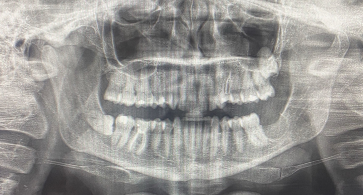
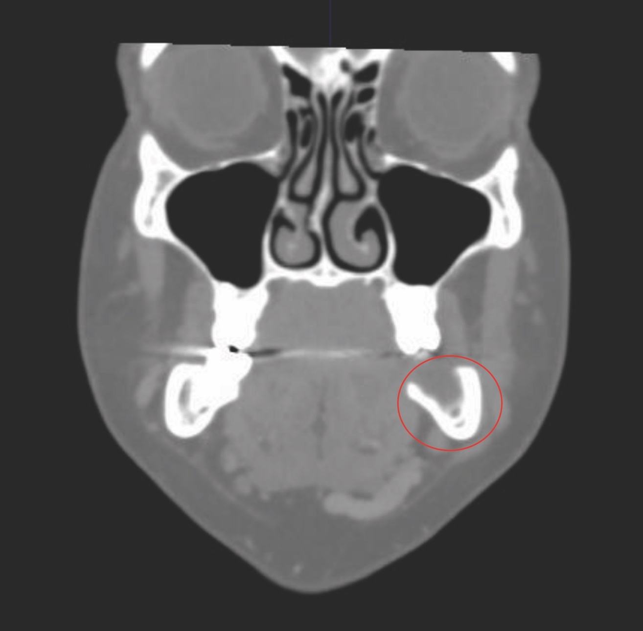
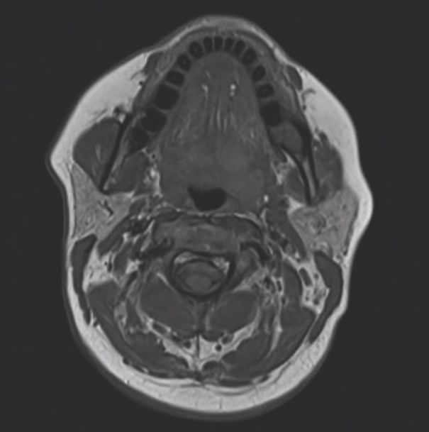
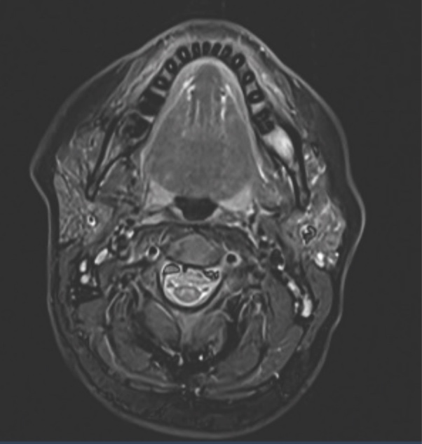
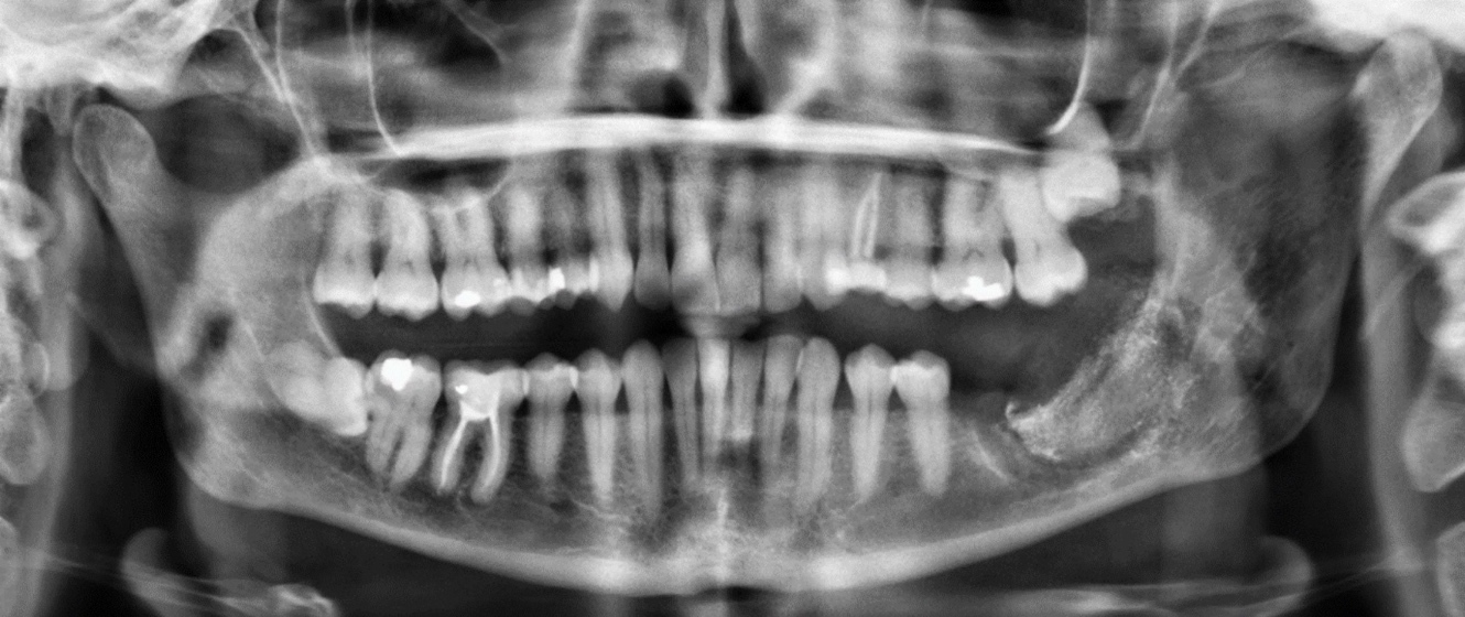
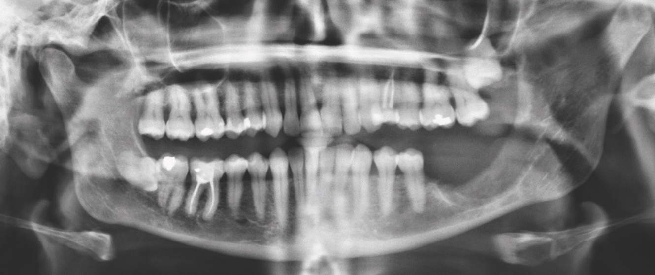
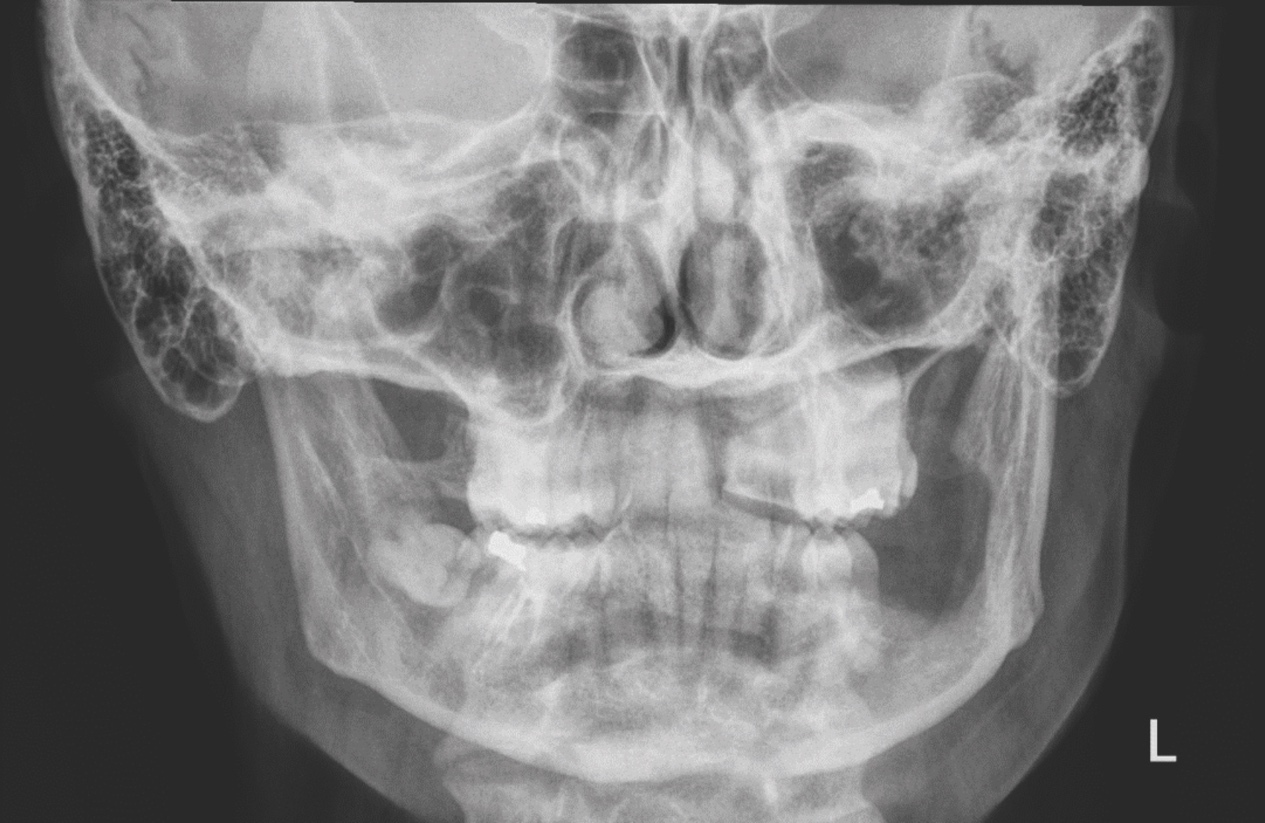
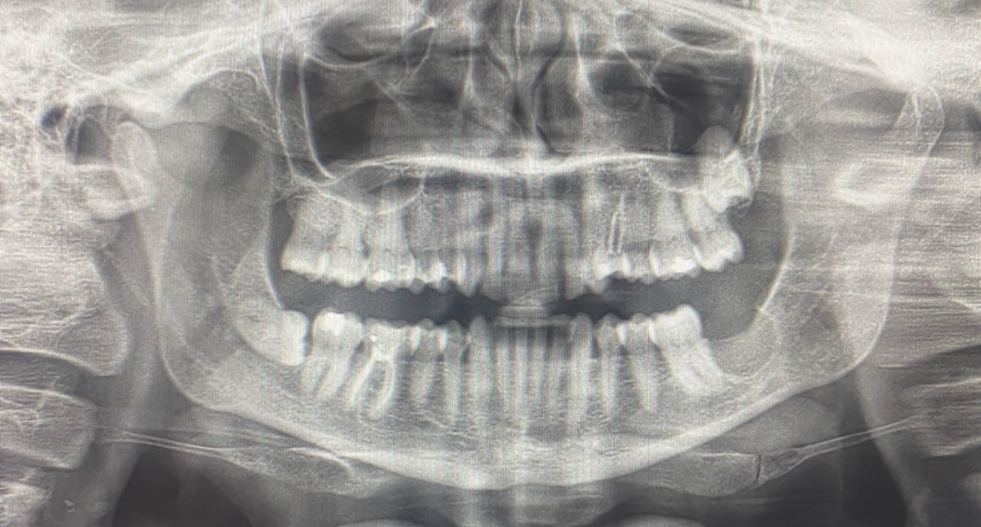
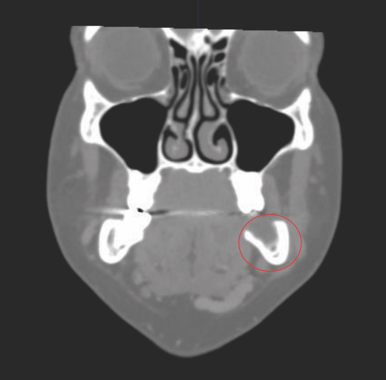
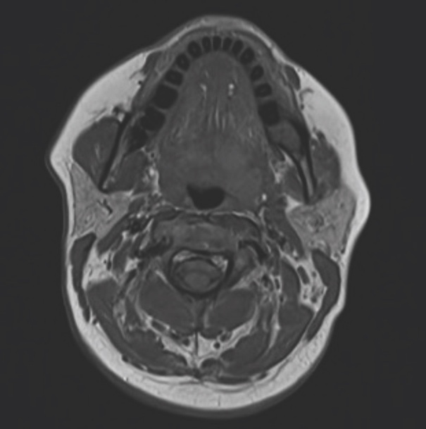
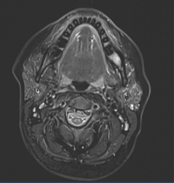
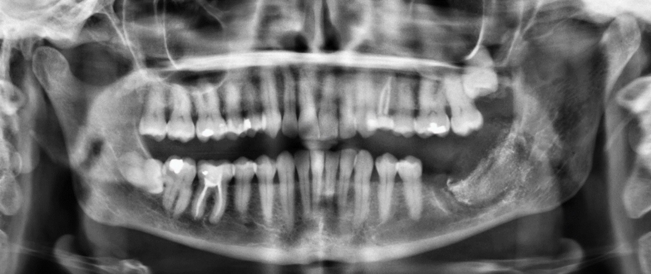
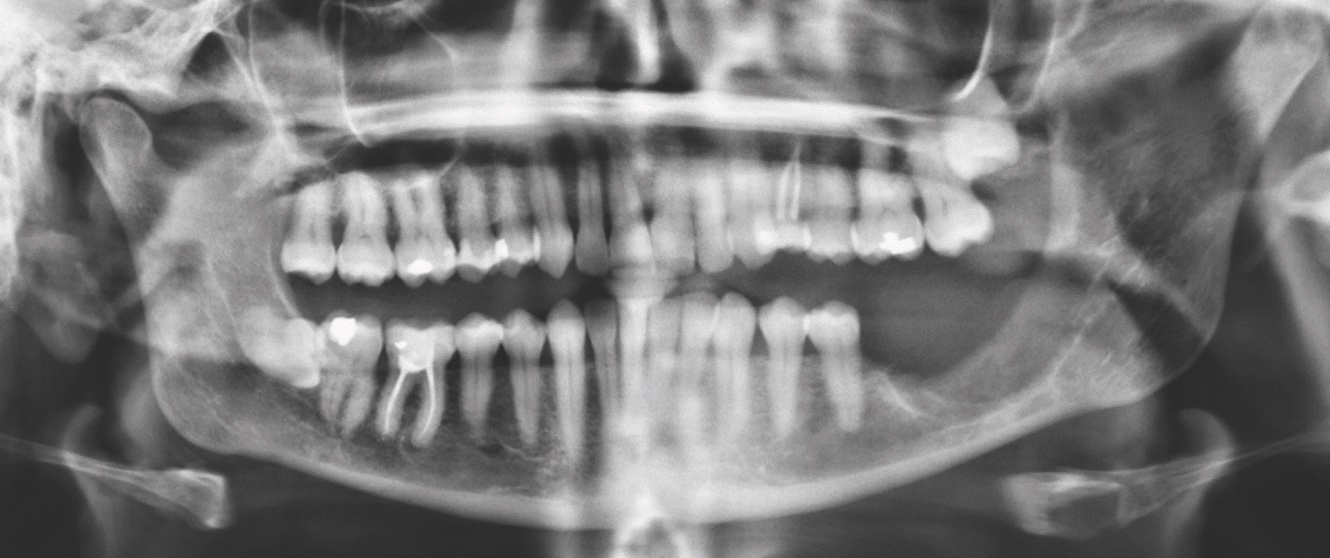
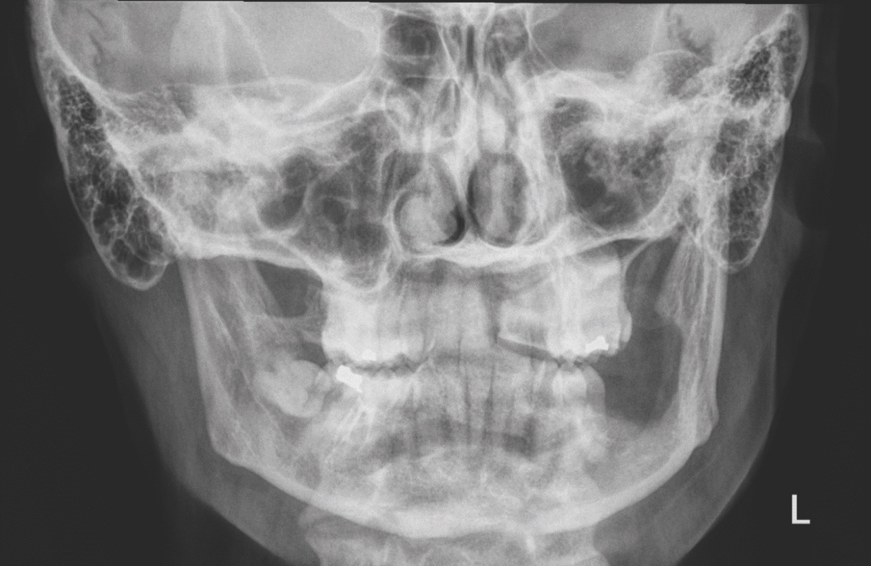
A lower left third molar had been surgically removed approximately three years earlier, and she described intermittent pain and tenderness in the same region during the intervening period. Earlier periapical and bitewing radiographs had been inconclusive. Subsequent cone-beam computed tomography (CBCT) demonstrated a well-defined unilocular radiolucent lesion involving the left mandibular body and angle, with associated cystic change on the lingual aspect, prompting biopsy (Figures 1 and 2). The lesion was closely associated with the inferior alveolar canal. Its well-circumscribed cystic appearance initially favoured an odontogenic lesion. Further cross-sectional imaging, including magnetic resonance imaging, was reviewed as part of the oncological assessment (Figure 3).
The initial differential diagnosis included a dentigerous cyst, odontogenic keratocyst and unicystic ameloblastoma. A primary intraosseous malignancy was considered less likely because of the well-defined radiographic margins and apparent relationship to the previous third molar site. However, the persistence of symptoms and subsequent atypical histological findings prompted further investigation.
Histopathological diagnosis
An incisional biopsy was initially reported as an inflamed dentigerous cyst with atypical features. Because the histological findings were unusual, the slides were referred to a specialist centre for review. This review supported a diagnosis of low-grade IMEC. Fluorescence in situ hybridisation demonstrated a MAML2 gene rearrangement, providing molecular support for the diagnosis.
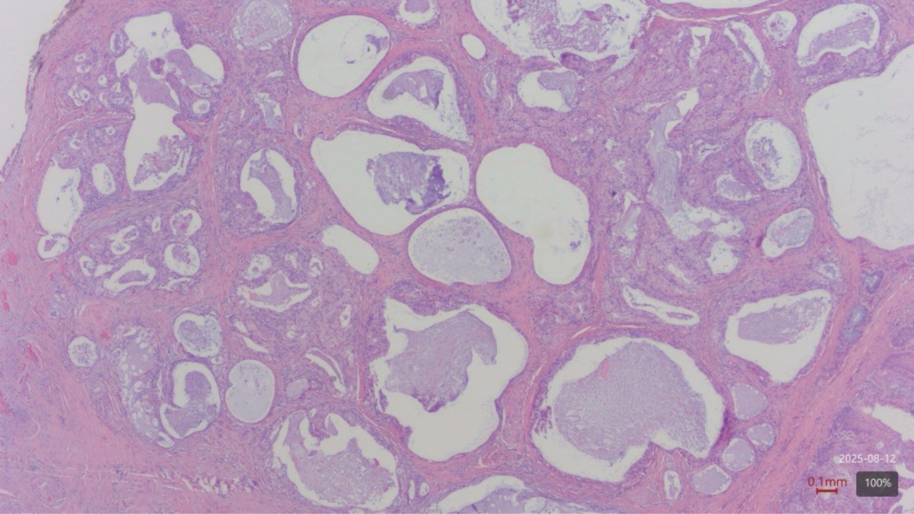
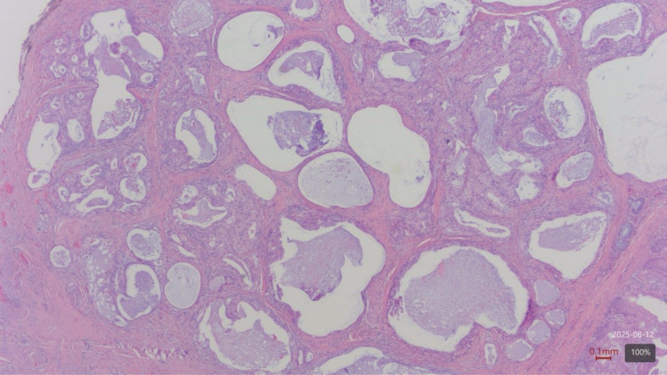
Final resection histology demonstrated an 8mm focus of low-grade mucoepidermoid carcinoma composed of multiple cystic spaces lined by intermediate, mucous and squamoid cells. The tumour was completely excised, with a closest margin of 3mm. A large nerve lay close to the tumour but was not involved, and no lymphovascular invasion was identified. The adjacent bone demonstrated cortical erosion, scarring fibrosis and remodelling. The pathological stage was pT1.
Multidisciplinary management
The case was discussed at a head and neck oncology multidisciplinary meeting. A marginal mandibulectomy was recommended to achieve oncological clearance while preserving mandibular continuity. Although the tumour was histologically low grade, enucleation or curettage was considered inappropriate because of the radiological extent of the lesion, its relationship to the lingual cortex and adjacent soft tissues, and the need to obtain reliable oncological margins.
Intraoperatively, the lesion was closely associated with the lingual soft tissues and floor of the mouth. The lingual and inferior alveolar nerves were inseparable from the lesion and were sacrificed to achieve adequate clearance. The lesion and adjacent bone were excised, the remaining cavity was curetted, and a Whitehead’s varnish pack was secured within the operative site.
Following review of the final histopathology, no additional treatment was recommended. Given the low-grade histology, pT1 stage, clear margins, and absence of lymphovascular or perineural invasion, continued surveillance was recommended without neck dissection or adjuvant radiotherapy.
Outcome and follow-up
Postoperatively, the patient experienced the anticipated sensory deficits affecting the ipsilateral tongue, lower lip and chin. She also developed reduced mouth opening, which was managed using a jaw-mobilisation device. Immediate and interval postoperative radiographs confirmed preservation of mandibular continuity (Figures 4 and 5).
At 12-month review, the patient remained clinically well, with no evidence of local or regional recurrence. Mouth opening was within normal limits. Altered sensation persisted in the left lower lip and chin; however, the patient was satisfied with the functional and overall outcome. Serial panoramic radiographs demonstrated no concerning abnormality, and continued surveillance was arranged. A subsequent CBCT approximately 15 months after surgery demonstrated postsurgical change in the left mandible, with no definite evidence of recurrent disease.
Discussion
Clinical and radiological presentation
IMEC most often presents with painless swelling, although pain, paraesthesia and tooth mobility may occur; some lesions are detected incidentally on imaging.6 The lesion may therefore enter the differential diagnosis of both benign and malignant jaw pathology. In the present case, intermittent symptoms and a radiolucent appearance at the site of a previous third molar extraction initially favoured an odontogenic origin. The panoramic radiograph obtained before the third molar extraction was not available for review.
Radiographically, IMEC may appear as a well-defined unilocular or multilocular radiolucency.7 This overlap with odontogenic cysts can delay diagnosis. A clinically important feature of this case was the discordance between the apparently cystic lesion and the atypical microscopic findings. Rather than accepting the initial benign interpretation, specialist pathological review was sought.
For the general dental practitioner, the more common differential diagnoses for a well-defined posterior mandibular radiolucency are odontogenic (Figure 1). A periapical lesion associated with a non-vital tooth may represent apical periodontitis, a periapical granuloma or a radicular cyst, while a similar lesion persisting at an extraction site may represent a residual cyst. A pericoronal radiolucency is more suggestive of a dentigerous cyst, although an odontogenic keratocyst or unicystic ameloblastoma may have a similar appearance. Tooth vitality, the relationship of the lesion to the crown or root apex, border definition, effects on adjacent teeth and the inferior alveolar canal, and evidence of expansion should therefore be assessed. Persistent symptoms, paraesthesia, progressive enlargement, or discordance between the clinical, radiological and histopathological findings should prompt specialist referral and tissue diagnosis.8
Diagnostic criteria and molecular confirmation
Diagnostic criteria reported by Browand and Waldron for central mucoepidermoid carcinoma include9:
-
intact cortical plates on computed tomography;
-
radiographic evidence of bony destruction;
-
exclusion of another primary tumour whose metastasis could histologically mimic the central tumour;
-
exclusion of an odontogenic tumour;
-
histopathological confirmation; and,
-
detectable intracellular mucin.
Histological grading categorises IMEC as low, intermediate or high grade (Figure 6). In this patient, the morphology supported a low-grade tumour. MAML2 rearrangement is a recurrent molecular alteration in mucoepidermoid carcinoma, often involving formation of a CRTC1–MAML2 fusion. Its detection supports mucoepidermoid differentiation when limited or inflamed biopsy material resembles the epithelial lining of an odontogenic cyst, although it does not independently establish an intraosseous site of origin.10
In this case, the radiological lesion was centred within the mandible and demonstrated bony destruction. Specialist histopathological review excluded an odontogenic neoplasm, while intracellular mucin and the MAML2 rearrangement supported mucoepidermoid differentiation. The definitive resection contained an 8mm focus of low-grade mucoepidermoid carcinoma in mucosa containing minor salivary glands, with cortical erosion and remodelling. The diagnosis was based on integrated assessment of the original clinical, radiological, biopsy, molecular and definitive histopathological findings.
Management and prognosis
Complete surgical excision is the principal treatment. Conservative procedures such as marsupialisation and enucleation have been associated with recurrence rates of approximately 40%, compared with approximately 13% following more definitive resection.11 The extent of surgery should nevertheless be individualised according to tumour grade, anatomical extent, and the ability to obtain clear margins. Segmental resection is required in many mandibular cases, while neck dissection and adjuvant radiotherapy are generally reserved for high-grade or otherwise adverse disease.1,6 Reported prognosis is influenced by histological grade, regional metastasis and the adequacy of surgical treatment.4 Five-year survival has been reported to approach 95% for low-grade lesions, but may be as low as 40% for high-grade disease.6 The low-grade histology and clear margins in this case supported surveillance rather than further treatment. Long-term follow-up remains important because the rarity of IMEC limits the available evidence regarding recurrence patterns and optimal surveillance.
Conclusion
IMEC is an uncommon malignant jaw tumour that can closely resemble a benign odontogenic cyst. This case demonstrates the importance of recognising discordance between clinical, radiological and histopathological findings. Specialist pathological review and molecular testing may prevent misdiagnosis, while multidisciplinary assessment enables treatment to be tailored to tumour grade and anatomical extent. Complete excision with clear margins and long-term surveillance remains central to management.
Learning points
-
A persistent or enlarging posterior mandibular radiolucency should not automatically be assumed to represent a benign odontogenic cyst;
-
tooth vitality, the lesion’s relationship to adjacent teeth and the inferior alveolar canal, and the presence of paraesthesia or cortical expansion should guide assessment and referral;
-
discordance between the clinical, radiological and histopathological findings should prompt specialist pathological review and consideration of molecular testing; and,
-
confirmed IMEC requires multidisciplinary management, complete excision where feasible, and long-term surveillance
Acknowledgements
The authors thank the oral and maxillofacial surgery, head and neck oncology, pathology, nursing and allied healthcare teams involved in the patient’s care. The authors also thank the patient for consenting to the publication of this case.
Funding
The authors received no funding for this work.
Conflicts of interest
The authors declare no conflicts of interest.
Patient consent
Written informed consent for publication was obtained from the patient.
Data availability
All data relevant to this case are included within the article.
Generative AI was used solely to assist with language editing, manuscript organisation and formatting. All clinical information, interpretations, references and final wording were reviewed and verified by the authors.