

Introduction
Supernumerary teeth are additional to the normal complement of 20 primary teeth and 32 permanent teeth. The presence of additional teeth can be referred to as ‘hyperdontia’, and supernumerary teeth can occur in 0.3-0.8% of primary dentitions and 1.2-3.5% of permanent dentitions.1 There may be under-reporting of primary dentition supernumerary teeth if they have erupted in an acceptable alignment or have exfoliated early. Supernumerary teeth are usually diagnosed at a single point in time and are referred to as non-sequential. In contrast, metachronous supernumerary teeth may develop and be identified at multiple time points throughout a patient’s lifetime.
In the permanent dentition, supernumeraries have been reported to be more common in males.1,2 This sex-related pattern is not observed in primary teeth. Patients who develop primary supernumerary teeth may exhibit a higher likelihood of presenting with this anomaly in the permanent dentition.3
Supernumerary teeth can be categorised by shape, including conical, tuberculate, supplemental and odontomas.4 Conical supernumeraries often exhibit a ‘peg-shaped’ appearance and typically manifest palatal to, or erupted between, the maxillary central incisors, often referred to as a mesiodens. Tuberculate supernumerary teeth are often located in the maxillary midline, are barrel-shaped, generally larger in size, and display incomplete root formation.4 The term ‘supplemental’ is used to describe well-formed additional teeth, commonly found in the upper lateral incisor region and also the premolar or molar region.1 Odontomas include ‘compound odontomas’ for tooth-like structures and ‘complex odontomas,’ for hamartomatous tooth-like formations.1
Supernumerary teeth can present as single or multiple teeth, unilateral or bilateral. Although supernumerary teeth can be located in any region of the maxilla or mandible, they are most commonly located in the premaxilla.2,3,5–7 The second most common location for multiple supernumeraries is the mandibular premolar region.5,6 Supernumerary teeth in the premolar region account for 8-10% of cases, with 75% presenting in the mandibular arch.5,6 Among supernumerary teeth in the mandibular premolar region, 75% are reported to be unerupted and asymptomatic.8 The mandibular premolar region has been reported as the most common site for multiple supernumeraries in non-syndromic patients.9 Yousof found that 44.8% of non-syndromic multiple supernumerary teeth occurred within the mandibular premolar area.9 Erupted supernumerary teeth can be identified through clinical examination. Unerupted supernumerary teeth are normally identified by radiographic examination.
Various theories have been proposed to explain the aetiology of supernumerary tooth development. The atavism theory, which proposes a connection to the evolution of the dentition, has been discredited by Primosch10 due to the high incidence of single supernumerary teeth and their ectopic position. Another theory, dichotomy, postulates that the tooth bud splits in two.11 Hyperactivity of the dental lamina is now the widely accepted aetiology of excessive tooth development.7 Primosch theorised that supplemental supernumeraries develop from an accessory tooth bud,7 and more poorly formed supernumerary teeth develop from the epithelial remnants of the hyperactive dental lamina.7
A positive family history is often observed with supernumerary teeth.5 There is no simple genetic pattern that has been identified, and the complex interactions of genetic/environmental interfaces have led to theories of the influence of environmental factors and the possibility of sporadic genetic mutations.12
Supernumerary teeth can adopt various orientations, either remaining impacted or erupting into the oral cavity. Studies by Stafne5 and Brook,13 have reported that around 25% of permanent supernumerary teeth have the potential to erupt.
Supernumerary teeth can contribute to delayed eruption of permanent teeth. Non-eruption of permanent central incisors has been reported to be associated with supernumerary teeth in 26-57% of cases.7 Supernumerary teeth can also result in median diastemas, rotations, periodontal defects or crowding, and have also been associated with dilaceration, abnormal permanent root development, cystic formation,1,6 and resorption of adjacent teeth with pulpal necrosis.12 During orthodontic treatment, unerupted supernumerary teeth can prevent space closure.13
Disorders that have been reported to exhibit an increased prevalence of supernumerary teeth include: cleft lip and palate (CLP); Gardner’s syndrome (GS); and, cleidocranial dysplasia (CCD).10,14,15 Multiple supernumerary formation is strongly linked to GS and CCD. Other less common syndromes associated with supernumerary teeth are: Fabry Anderson syndrome16; Ellis-Van Creveld syndrome17; Ehlers-Danlos syndrome18; incontinentia pigmenti; and, trico-rhino-phalangeal syndrome.19
CCD is a rare autosomal dominant disorder, characterised by skeletal and dental anomalies. Classic CCD presents with a triad of symptoms considered to be pathognomonic of the condition: absent or hypoplastic clavicles; multiple supernumerary teeth; and, open cranial sutures.20 Patients with CCD often present with delayed eruption of permanent teeth, root abnormalities, ectopic teeth, prolonged retention of deciduous teeth, supernumerary teeth, and an underdeveloped maxilla resulting in a Class III malocclusion.19
Case report
This patient first attended the Oral Surgery Department of Royal School of Dentistry Belfast for removal of a compound odontome in the right mandibular premolar region. This was an incidental finding discovered during assessment for orthodontic treatment (Figure 1). There was no evidence at that time of any other supernumerary teeth elsewhere in the jaws. This compound odontome tooth was surgically removed at that time following the informed consent process. The patient subsequently completed orthodontic treatment.

Eight years later, at the age of 21, the patient was referred to the Oral Surgery Department for a second time due to the incidental finding of another supernumerary tooth on the contralateral side (Figure 2). The patient reported no relevant medical or genetic conditions. There was no reported family history of supernumerary teeth.

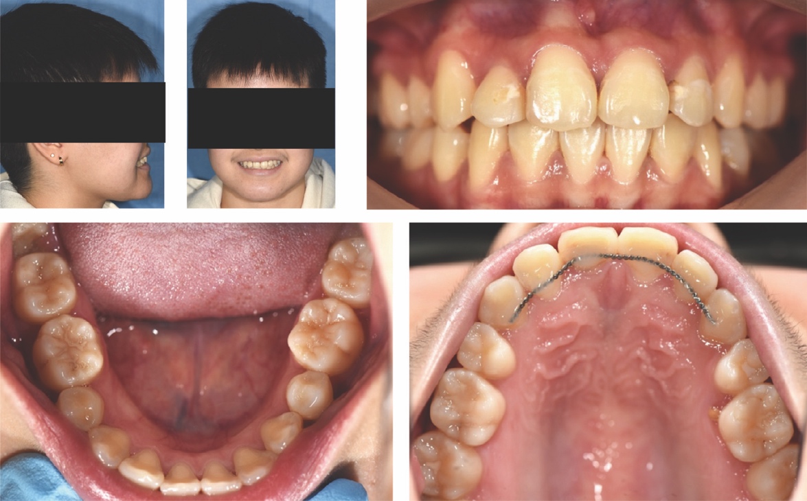
Clinical examination at age 21 years revealed a well-aligned dentition with a maxillary bonded retainer in place (Figure 2). No symptoms were reported related to the supernumerary teeth. None of the teeth were tender to percussion and all adjacent teeth exhibited a positive response to ethyl chloride. There was a palpable lingual bulge in the alveolar region of the lower left second premolar and first permanent molar.
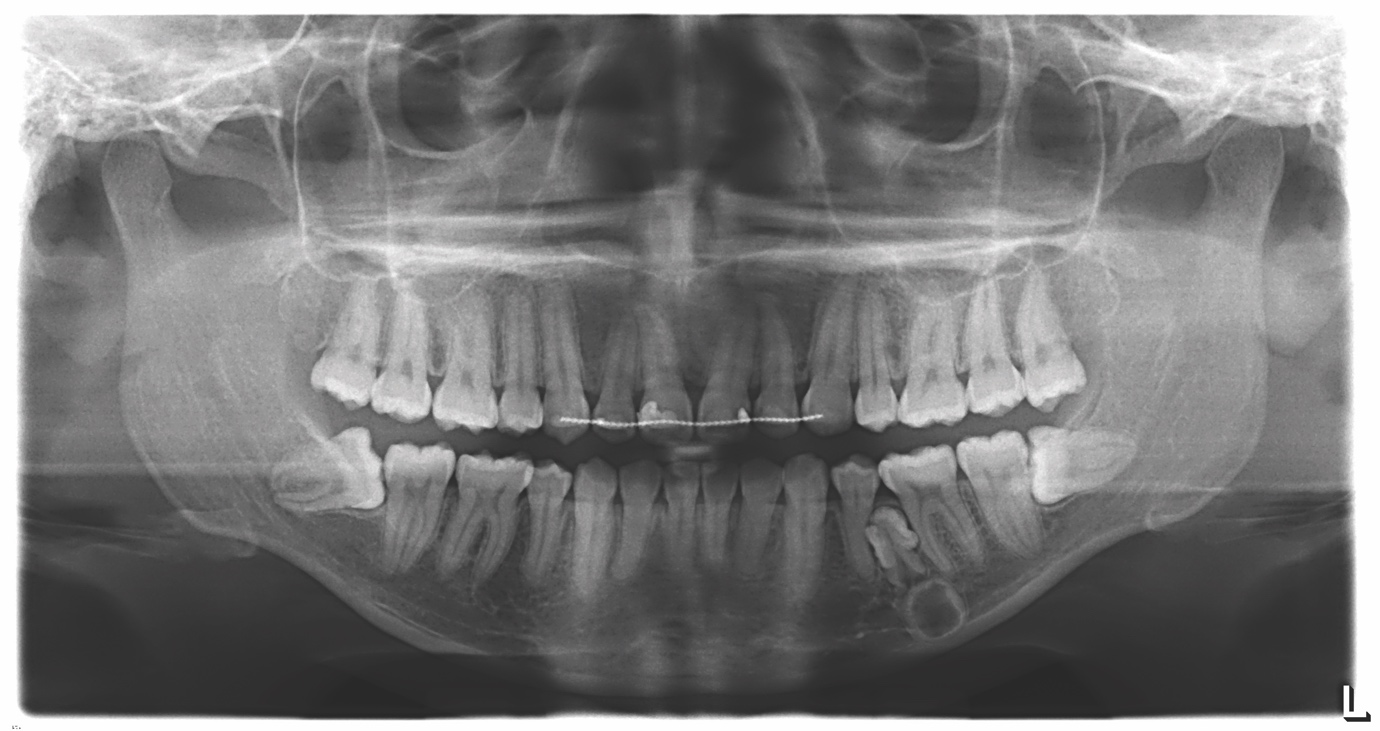
The panoramic radiograph indicated the presence of a radiopaque lesion and a radiolucent lesion in the left mandibular premolar region (Figure 3). A cone-beam computed tomography scan was recorded to further characterise the lesion and surrounding area to aid surgical planning (Figure 4).


A maxillofacial radiology report confirmed the presence of fused supernumerary teeth with abnormal crown forms, with an inferiorly located mixed density lesion. The circular mixed density lesion had a radiodense periphery and a radiolucent core superimposed by the inferior alveolar canal. The follicle of the fused supernumeraries and that of the mixed density lesion were confluent. The mixed density lesion was consistent with an invaginated (dilated) odontome. Minor root resorption was reported into the outer dentine of the lower left premolar and lower left first molar.
The patient was happy with the outcome of their previous orthodontic treatment and no further orthodontic treatment was planned. The risks and benefits of surgical removal of the supernumerary teeth were discussed with the patient, including consideration of the proximity of the roots of adjacent teeth, and the inferior alveolar canal. It was agreed that a further radiographic reassessment would be carried out in 18 months to monitor for the possibility of cystic change or resorption. The possibility of further supernumerary development was discussed with the patient.
Discussion
We have described the case of metachronous development of supernumerary teeth and odontome in the right mandibular premolar region, eight years following removal of a supernumerary in the right mandibular premolar region.
Multiple supernumerary teeth can be associated with a developmental genetic aetiology and associated syndromes such as GS and CDD.10,15 While supernumerary teeth are a common clinical phenomenon, metachronous supernumerary teeth are rare.21 Non-syndromic multiple supernumerary teeth have been reported to have a predilection for the mandibular premolar region.9 A review of the literature did not reveal any case reports of metachronous supernumerary formation in syndromic patients.
Multiple supernumerary teeth are defined as more than three or five. The reported prevalence of multiple supernumeraries varies within the literature, with some studies reporting less than 1% of cases while others indicate a prevalence of 14% in certain populations.6,7
The timing of supernumerary development in this case is atypical, and presented at 21 years. It is generally agreed that supernumeraries develop later than the normal dentition, attributed to delayed root development observed radiographically. It is estimated that supernumerary premolars exhibit a delayed development of 7-11 years compared to normal premolars.22 Case studies documenting late developing supernumerary premolars consistently indicate that these supernumeraries tend to appear later than supernumeraries in other locations.23 Oehlers reported continued supernumerary premolar root growth in a 23-year-old man. Due to their delayed development, supernumerary premolars have been postulated to be part of a third series of teeth, supporting the post-permanent dentition theory as an explanation for this phenomenon.24
Treatment planning requires consideration of various tooth factors, including angulation and proximity to the cortical bone, inferior alveolar nerve, mental nerve, blood vessels, and neighbouring teeth. Moreover, management is significantly influenced by patient-specific factors, including their capacity to withstand interventions, aesthetic considerations, the current symptoms and medical background.
Two approaches are available for the management of supernumerary teeth. The first option is proactive removal. Theoretically, this approach offers the advantage of removing further risk of root resorption of adjacent teeth. Supernumeraries do not usually cause resorption, although if they develop roots, they may have the eruptive potential to cause resorption of adjacent teeth.7 Removal of a supernumerary may reduce the risk of cystic formation and ankylosis.6,7 Timely removal also can harness the eruptive potential of obstructed teeth and has been described to provide an improved prognosis.7 The bone quality in younger patients may make removal easier, although this must be weighed against the increased likelihood of requiring a general anaesthetic. This was the approach taken to manage the right mandibular supernumerary at age 13 in this patient to facilitate orthodontic tooth movement and to reduce the risk of root resorption of adjacent teeth.
An alternative approach involves radiographic monitoring in the absence of symptoms or planned orthodontic treatment. The advantages of radiographic monitoring include the lack of postoperative pain and discomfort, infection, bruising, swelling, and avoidance of potential nerve trauma. Avoidance of a general anaesthetic also removes risk of mortality or morbidity. Leaving the supernumerary in situ can remove the risk of post-surgical permanent numbness or devitalisation of adjacent teeth. If the supernumerary is located near immature roots, delaying treatment until these have fully developed can minimise potential growth disruption.25 Garvey et al. supported this approach in patients with the absence of symptoms or pathology, where all permanent teeth have erupted unimpeded and where no orthodontics is planned, and that approach was adopted in this case. Radiographic evaluation prior to orthodontic treatment – including in cases of retreatment – remains essential. Clinicians should maintain an index of awareness that additional supernumerary teeth can emerge over time even if this is exceedingly rare.21
Conclusion
This case report demonstrates an uncommon presentation of supernumerary teeth. It is unusual for multiple supernumerary teeth to present in the absence of an associated syndrome. Nevertheless, there are previous reports of supernumerary teeth developing in the mandibular premolar region in late adolescence and early adulthood. Clinicians should make patients aware of the possibility of the future development of additional supernumerary teeth. Supernumerary teeth can often be managed conservatively in the absence of complications, with appropriate longitudinal radiographic examination. This is particularly important in patients who are subsequently considering orthodontic treatment. Careful consideration should be given to their removal in relation to the risks to adjacent structures and the benefits of removal. In cases with additional clinical features, consideration should be given to a genetic aetiology.